the separate parts · unequal support
What each KLOW stack ingredient changed in studies
Researchers chose KPV, GHK-Cu, BPC-157 and TB-500 for possible repair effects. Skin creams and animal tests showed some changes with separate ingredients. Those findings don’t establish better healing from the KLOW peptide blend for you.
What the first KLOW stack makers wanted to add together
The first mixers hoped to join several repair effects. They chose ingredients studied for inflammation, tissue strength and blood supply. The combined mixture became known as the KLOW stack. Putting the parts together hasn't shown better healing for you.
Some parts helped injured animals, and others changed lab-grown cells. Cream research also found changes in people's skin with one ingredient. The studies didn't test all four together in injured people. Your injury benefit remains unknown because studies of different animals, cells and skin creams don't show what happens when someone combines all the ingredients into a single mixture for repair.
What KPV reduced, and what remains unknown for bowel trouble
Human bowel cells in dishes produced less of the substances driving inflammation after KPV [3]. The reduction was measured in those treated cells. Researchers used 10 nanomolar KPV, stating the amount of dissolved KPV for each liter of test liquid [3]. That dilute liquid changed cells, without showing a treatment benefit for you.
KPV contains three parts of the kind that make proteins. The ingredient comes from a larger hormone, a substance carrying messages around the body. Bowel cells absorbed KPV from liquid surrounding them. Absorption doesn't by itself show a treatment benefit for your bowel disease.
Researchers reported about 160 micromolar in the absorption test [3]. Micromolar measures how much KPV was dissolved in each liter. The test concerned how readily cells absorbed KPV, rather than bowel pain or healing. Inflamed bowel cells absorbed KPV more readily, but weren't people receiving treatment.
Active immune-system cells also absorbed KPV. After treatment, the cells produced less of the substances linked to swelling and damage. Reducing those substances could explain interest in bowel illness. A completed human trial hasn't shown that these effects relieve your illness.
Mice with inflamed bowels improved after receiving KPV by mouth. A KPV gel also reduced swelling and helped animal wounds [12]. Those experiments tested the ingredient separately. They don't show extra benefit for you from the other mixture parts.
KPV doesn't have approval to treat people or bowel disease. Early human checks concerned delivery, without establishing relief from bowel illness. You aren't told whether people swallowed the ingredient, received a shot or used another method. Neither relief nor a safe treatment amount was established by those checks.
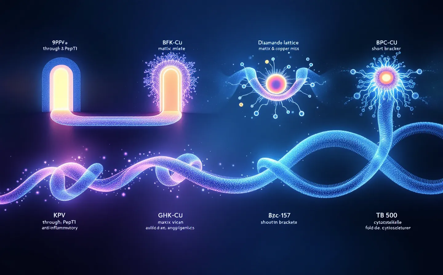
What GHK-Cu improved on skin, rather than inside an injury
With GHK-Cu cream, more collagen was produced in women's skin. Collagen is a protein your skin uses for strength. More collagen appeared in 70% with GHK-Cu, 50% with vitamin C and 40% with retinoic acid, derived from vitamin A and applied to skin [4]. Those figures describe women whose skin changed.
The cream research also found better skin texture and finer lines. The women received creams, rather than shots. Putting cream on skin differs from injecting an ingredient. You can't treat the cream findings as proof of injury repair.
Loren Pickart first separated GHK-Cu from blood in 1973. A 20-year-old had a reported level of 200 nanograms per milliliter in blood; someone aged 60 had 80 nanograms per milliliter [4]. Nanograms count tiny weights in a measured amount of blood. You can't assume that GHK-Cu restores youth because older blood contains less.
Copper Tripeptide-1 means copper joined to three protein parts; it's another name for GHK-Cu. This ingredient has the largest bottle weight. A 2018 study found that cells changed their use of genes, their instructions for making proteins [5]. The use had to rise or fall by at least 50% to be counted.
Some changed genes concerned building tissue and removing worn proteins. Others concerned repairing DNA, which carries inherited instructions within your cells. Protection against damage also changed. None of those cell findings measured a person's injury recovery.
Copper helps collagen and stretchy proteins join together in tissue. GHK-Cu carries copper that cells can use for those jobs. Both skin and tendons need collagen for strength. Your benefit from mixing isn't established by those separate effects.
Related copper-carrying pieces are released when another tissue protein breaks down. Those pieces encouraged new vessels when tested in animals or on cells [9]. Researchers weren't testing the complete mixture. Vessel growth alone hasn't settled either your risks or your recovery.
What BPC-157 helped in rats, and what its human check missed
BPC-157 was first developed for inflammatory bowel disease, which causes swollen, damaged bowels. An earlier research name was PL 14736. Later work tested injuries in tendons, ligaments, muscles, bowels and blood vessels. Most repair findings came from animals, without establishing your own recovery.
BPC-157 belly shots helped rats' tendons regain movement and strength [2]. Researchers had cut through their Achilles tendons. Collagen, the protein that strengthens tendons, was better arranged afterward. A better rat tendon doesn't prove that your tendon would mend.
All three daily amounts showed repair effects: 10 millionths, 10 billionths and 10 trillionths of a gram per kilogram of the rat’s weight. Kilograms measure the rats' body weight. The smallest amount helped, but this account doesn't establish equal help. These weren't amounts tested for your treatment.
Researchers linked BPC-157 effects to new blood vessels [10]. A protein called growth receptor 2 helps start vessel growth. The ingredient also changed how vessels widened and tightened [10]. Treated tendon cells had more places than untreated cells for growth hormone, a messenger for growth, to attach.
Some vessels still widened despite a chemical used to stop widening. That suggests the ingredient affected vessel width another way. The researchers were testing vessel behavior. Those tests didn't show a person's injury healing or pain easing.
The 2025 safety check involved a 58-year-old man and a 68-year-old woman [6]. They received 10 mg on day 1 and 20 mg on day 2 through a vein. Researchers used 250 cc of salt water, roughly a cup, to carry the ingredient. Neither reported harm; heart, kidney, liver, blood-sugar and thyroid tests stayed unchanged, with thyroid tests checking the neck gland that helps control energy use.
Only two adults were checked, without establishing safety for you. FDA put BPC-157 in Category 2 for serious safety concerns in September 2023. Someone seeking permission for pharmacy-made medicines later withdrew the review request. FDA's page current as of April 22, 2026 lists the ingredient outside Category 2 because the request for review was withdrawn.
The withdrawn request wasn't a finding that the ingredient is safe. It wasn't treatment approval either. FDA hasn't explained whether pharmacies can make the ingredient after the request ended. Your benefit and risks are still unproved.
What TB-500 hasn’t shown despite results with the full protein
The strongest wound findings used thymosin beta-4, a full protein your body makes. Malinda's 1999 study found 42% more wound coverage in rats at 4 days and 61% at 7 days [1]. Wound edges drew together at least 11% more by day 7. The wounds formed vessels and gained collagen, a protein keeping tissue strong.
Researchers added the full protein to skin cells in dishes, using 10 trillionths of a gram. Cell movement increased 2–3-fold, or between twice and three times the earlier movement. That added weight doesn't state the liquid's strength. Moving cells don't establish healed wounds in people.
Thymosin beta-4, the full body protein, has a 43-amino-acid chain, with forty-three linked protein parts. TB-500 is made from only part of the full protein. The shorter ingredient is linked to movement of cells during repair. Movement alone doesn't show that your wound would heal better.
The full protein also activates certain repair cells. The short piece hasn't shown those extra effects. TB-500 can behave differently from the full protein. You can't assume TB-500 heals because thymosin beta-4 helped rats' wounds.
Thymosin beta-4, the full protein, increased a substance encouraging blood-vessel growth [11]. A 2021 review considered possible hair-growth effects [8]. A 2026 Sports Medicine review included TB-500 among unapproved substances [7]. Animal repair findings gave reasons for interest, but few studies had checked safety in people and serious harm remained possible.
Sports rules create another limit if testing applies to you. Under WADA rules, thymosin beta-4, the full body protein, is banned all year. TB-500 is taken from that protein, so the sports restriction carries over. Calling use research doesn't cancel the sports drug ban.
What combining the four repair ideas still needs to show
Separate ingredients showed separate effects, without proof of a better mixture. KPV reduced inflammation-causing substances, and GHK-Cu helped skin and tissue-building cells. BPC-157 affected blood vessels, while TB-500 has links to cell movement. Combining hasn't shown that your injury mends better because of those results.
BPC-157 has many animal repair findings; GHK-Cu has findings from human creams. The full protein behind another ingredient has animal wound results. TB-500 has less support, and early human KPV work concerned delivery. You still lack proof of relief, and you aren't given a clear account of how the ingredient was delivered to people.
You lack a healing test of the complete mixture against fewer ingredients. Researchers also haven't fully checked how dissolved ingredients affect one another. KLOW effects describes accounts from users and possible harms raised by separate studies. Why users felt relief or discomfort hasn't been checked in a trial.